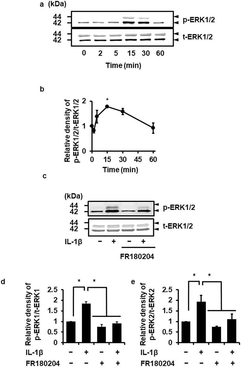
Fig 3. IL-1β-induced ERK1/2 phosphorylation and its inhibition by an ERK1/2 inhibitor.
In dermal fibroblasts treated with 100 pM IL-1β for 0–60 min, ERK1/2 phosphorylation (p-ERK1/2) was observed in a time-dependent manner (a, b). IL-1β had no effect on total ERK1/2 (t-ERK1/2) expression (a). In cells pretreated without (control) or with the ERK1/2 inhibitor FR180204 (25 μM) for 1 h, IL-1β-induced ERK1/2 phosphorylation was attenuated (c, d). Representative results (a, c) and the relative density of ERK1/2 phosphorylation compared with the results at 0 time (b) or the control (d) are illustrated. Values are expressed as the mean ± SE of 3 independent experiments (b, d). *P < 0.05.

