
Fig. 2. Nrf2-dependent mitigation of hyperoxia (O2)-induced pulmonary histopathology by dietary glucoraphanin (SBE).
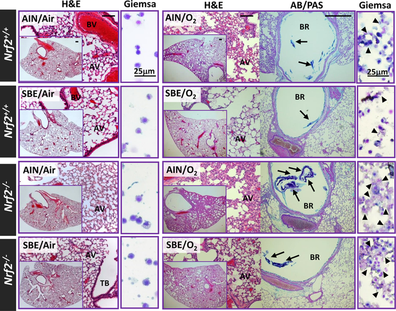
Representative light photomicrographs of lung tissue sections stained with H&E and AB/PAS or cytocentrifuged BAL cells stained with Giemsa after 14 days of normal (AIN) or standardized broccoli extract-containing diet (SBE) followed by 72 h exposure to normoxia (room air) or O2. AV, alveoli; BR, bronchi; BV, blood vessel; TB, terminal bronchiole. Arrows=secreted mucus stained. Arrow heads=lysed cells. Bars (unlabeled) = 100 μm.