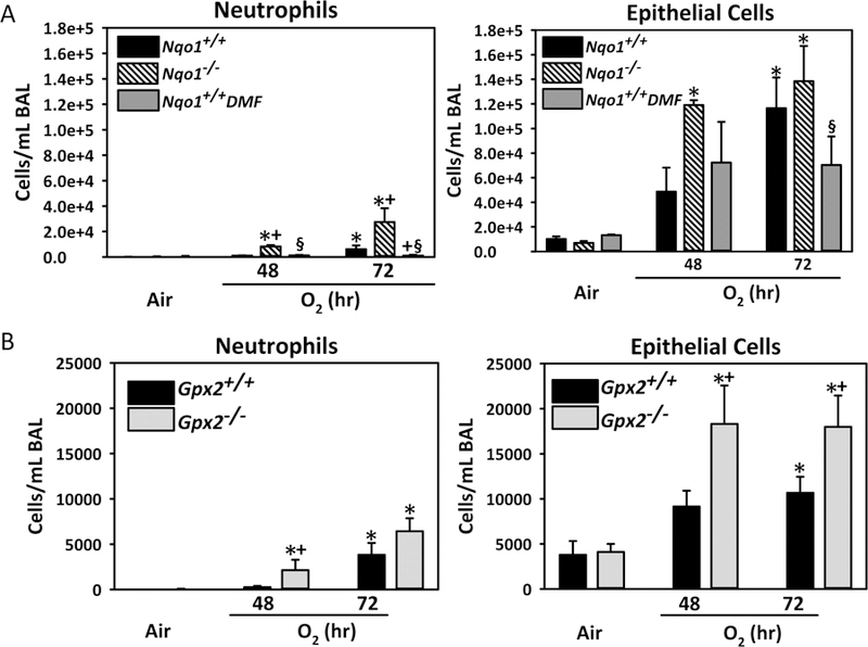
Fig. 8. Role of Sulforaphane-responsive antioxidant enzymes in hyperoxia-induced lung injury.

(A) Numbers of neutrophils and epithelial cells found in bronchopulmonary lavage (BAL) fluid from wild-type (Nqo1+/+), dimethyl fumarate (DMF)-fed wild-type (Nqo1+/+DMF), and Nqo1−/− mice after exposure to air and O2 (48–72 h). Data presented as group mean ± SE (n = 3/group for air, n = 5/group for 48 h O2, n = 7/group for 72 h O2). (B). Numbers of neutrophils and epithelial cells found in BAL fluid from Gpx2+/+ and Gpx2−/− mice after exposure to air and O2 (48–72 h). Data presented as group mean ± SE (n = 3/group for air, n = 6/group for 48 h O2, n = 10/group for 72 h O2). Two-way ANOVA used for statistical analyses. *, P < 0.05 vs. genotype and air controls. +, P < 0.05 vs. exposure-matched wild-type mice. §, P < 0.05 vs. exposure-matched Nqo1−/− mice.
