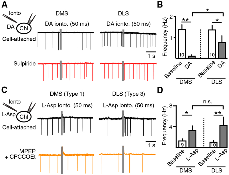
Figure 4. Post-synaptic Responses of DMS and DLS ChIs in Response to Iontophoretic Application of Dopamine or L-aspartate.

(A) Sample traces of cell-attached recordings from DMS and DLS ChIs in response to iontophoretic application of dopamine (1 M, 50 ms).
(B) Quantification of the dopamine-induced decrease in firing in DMS and DLS ChIs.
(C) Sample traces of cell-attached recordings from a type 1 DMS ChI and a type 3 DLS ChI in response to iontophoretic application of L-aspartate (200 mM, 50 ms).
(D) Quantification of the L-aspartate-induced increase in firing in DMS and DLS ChIs.
Error bars represent SEM. *p < 0.05, **p < 0.01.
