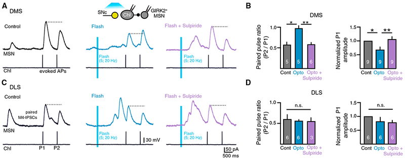
Figure 7. Dopamine Inputs Inhibit Acetylcholine Release at Muscarinic Synapses Only in the DMS.

(A) Paired recordings made from ChI-dMSN pairs in the DMS. A pair of evoked action potentials in control condition (black) and paired IPSCs evoked 1.5 s after photostimulation of dopamine terminals (flash, blue) and in the presence of sulpiride (500 nM, flash + sulpiride, purple). ChIs hyperpolarized to prevent firing except when triggered. Inter-pulse interval P1 to P2 is 750 ms.
(B) Left: quantification of paired-pulse ratios of paired IPSCs for the paired recordings represented in (A). Right: normalized amplitude of paired M4-IPSCs under control conditions (black), following photoactivation of SNc dopamine terminals (flash, blue) or following photoactivation of SNc dopamine terminals in the presence of sulpiride (500 nM) (flash + sulpiride, purple) from the DMS.
(C) Left: paired recordings are made from ChI-dMSN pairs in the DLS.
(D) Quantification of paired-pulse ratios of paired IPSCs for the paired recordings represented in (C). Right: normalized amplitude of paired M4-IPSCs under control conditions (black), following photoactivation of SNc dopamine terminals (flash, blue) or following photoactivation of SNc dopamine terminals in the presence of sulpiride (500 nM) (flash + sulpiride, purple) from the DLS.
Error bars represent SEM. *p < 0.05, **p < 0.01.
