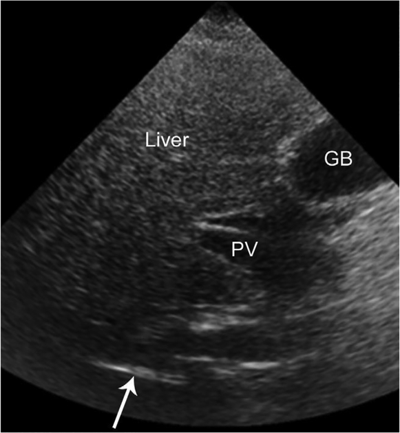
Figure 2.
Transverse US image of the liver in a 45-year-old man with fatty liver disease. The liver is hyperechoic in the near field (top half of image). Because of beam attenuation, the liver becomes progressively hypoechoic in the far field (bottom half of image). Note that only a portion of the right hemidiaphragm (arrow) is visible. Beam attenuation obscures the remainder of the hemidiaphragm. In addition, there is a paucity of intrahepatic structures. The portal vein (PV) is visible in the porta hepatis, but its intrahepatic branches are not evident. The gallbladder (GB) is incidentally noted.

