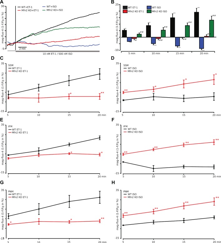
Figure 5.
ATP production following IP3-mediated SR Ca2+ release is impaired in Mfn2 KO mice. (A) Original confocal traces of ATP levels recorded in WT myocytes stimulated with 10 nM ET-1 (black) or 500 nM ISO (blue) and Mfn2 KO cardiac myocytes stimulated with ET-1 (red) or ISO (green), respectively. Mg2+-sensitive fluorescent dye mag-fluo-4 was used to monitor ATP levels inside cardiac myocytes. As free Mg2+ and ATP form complexes, increase in ATP concentration reduces free Mg2+ and thus mag-fluo-4 fluorescence. Traces are inverted for better understanding and presented as 1-F/F0. (B) Mean values of the experiment described in (A) in %. *p ≤ 0.05 compared to untreated WT; **p ≤ 0.01 compared to untreated WT; #p ≤ 0.05 compared to WT treated with ET-1 or ISO, respectively; ##p ≤ 0.01 compared to WT treated with ET-1 or ISO, respectively. (C–H) Subgroup analysis of subsarcolemmal (SSM, C,D), interfibrillar (IFM, E,F), and perinuclear (PNM, G,H) mitochondria in myocytes from WT (black) and Mfn2 KO (red) mice loaded with mag-fluo-4 as described in (A). The myocytes were treated with either ET-1 (left) or ISO (right). *p ≤ 0.05 compared to WT, **p ≤ 0.01 compared to WT.

