. 2019 Jul 15;8:e46683. doi: 10.7554/eLife.46683

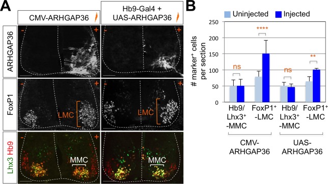
Figure 6. Expression of ARHGAP36 promotes LMC specification in developing chick spinal cord.

(A) ARHGAP36 constructs were injected and electroporated in chick neural tube and embryos (n = 8 ~ 15) were harvested 4 days post electroporation (four dpe). Ectopic expression of ARHGAP36 driven by CMV promoter in most injected cells induced robust expression of FoxP1+ LMC neurons (orange bracket) in ventral spinal cord but had no effect on MMC (Hb9+/Lhx3+) neurons (white bracket). Targeting the expression of ARHGAP36 specifically in motor neurons using Hb9-Gal4/UAS-ARHGAP36 system also lead to the robust induction of FoxP1+ LMC neurons (orange bracket) but had no effect on MMC (Hb9+/Lhx3+) neurons (white bracket). +, electroporated side; -, non-electroporated control side. Experiments were repeated independently at least three times. Scale bars: 100 μm. (B) Quantification of the number of FoxP1+ neurons and MMC (Hb9+/Lhx3+) neurons on the electroporated (+) and non-electroporated (-) sides of the spinal cord. Data are mean ± s.d. **p<0.001, ****p<0.00001; ns, non-significant (Student’s t-test). n = 6 ~ 20 independent images per each sample.

Figure 6—source data 1. Source data for Figure 6B.
DOI: 10.7554/eLife.46683.019

Figure 6.

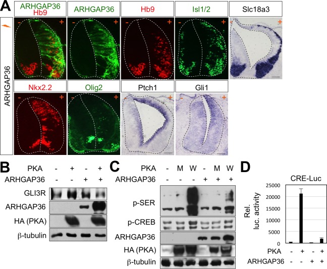
Figure 6—figure supplement 1. Activation of Shh pathway by ARHGAP36 expression in spinal cord.

Figure 6—figure supplement 1.

(A) In ovo electroporation of ARHGAP36 construct. ARHGAP36 construct was injected and electroporated in chick neural tube and embryos (n = 8 ~ 12) were harvested 3 days post electroporation (three dpe). Ectopic expression of ARHGAP36 induced expression of ventral spinal neuronal genes including motor neuron genes (Hb9, Isl1/2, Slc18a3), ventral progenitor genes (Nkx2.2 and Olig2) and downstream target genes of Shh pathway (Ptch1 and Gli1) as shown by ISH and IHC. +, electroporated side; -, non-electroporated control side. Experiments were repeated independently at least three times. Scale bars: 100 μm. (B) Western blot analysis showed that ARHGAP36 inhibits PKA activity. PKA expression resulted in producing Gli3R, repressor form of Gli3 (lane 2) in HEK293T cells. Co-expression of ARHGAP36 blocks the formation of Gli3R by PKA (lane 4). β-tubulin was used as a loading control. (C) ARHGAP36 inhibited the level of phosphorylation of putative PKA substrates shown by western blot with anti-p-SER antibody and the level of phospho-CREB (p-CREB), a known PKA target in NIH3T3 cells. W indicates PKA wild type and M indicates PKA kinase dead mutant (K73H). β-tubulin was used as a loading control. (D) Luciferase reporter assay in HEK293T cells. The activation of CRE-luc is directed by PKA-phosphorylated CREB and this activity was blunted by co-expression of ARHGAP36, indicating that ARHGAP36 inhibits the kinase activity of PKA. Data are mean ± s.d.
Figure 6—figure supplement 1—source data 1. Source data for Figure 6—figure supplement 1D.
DOI: 10.7554/eLife.46683.017
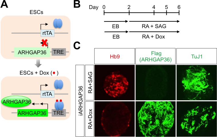
Figure 6—figure supplement 2. ARHGAP36 is not sufficient to induce MN differentiation in mESCs.

Figure 6—figure supplement 2.

(A) Schematic representation of inducible ARHGAP36-ESCs before and after treatment with Dox (Doxycycline). TRE, tetracycline response element; rtTA, reverse tetracycline transactivator. (B) Illustration of experimental design to differentiate ESCs to MNs. RA and SAG or RA and Dox were treated at day two and EBs were harvested at day six for further analyses. EB, embryoid body; RA, retinoic acid; SAG, Smoothened agonist. (C) IHC analyses in ESC-derived MNs cultured with RA +SAG or RA +Dox at differentiation day 6. Hb9 labels MNs and it was induced only by RA +SAG. Treatment of RA +Dox induced ARHGAP36 and pan-neuronal marker TuJ1, but not MN marker Hb9.