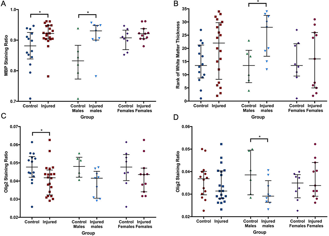
Figure 7. Quantitative immunohistochemistry.
Within the internal capsule, MBP (A) staining ratio was significantly greater and less variable in the injured group compared to the control group. This was particularly evident in male animals. The thickness of the corpus callosum and three areas of the internal capsule at the base of consecutive sulci were then measured, and a summary score based on the ranked weight-adjusted thickness of all four areas (B) suggested thinning of the white matter in injured males. Olig2 staining intensity was decreased in the corpus callosum (C) of injured animals compared to control animals. Olig2 staining ratio was also lower in injured males compared to control males within the internal capsule (D). * denotes p<0.05.

