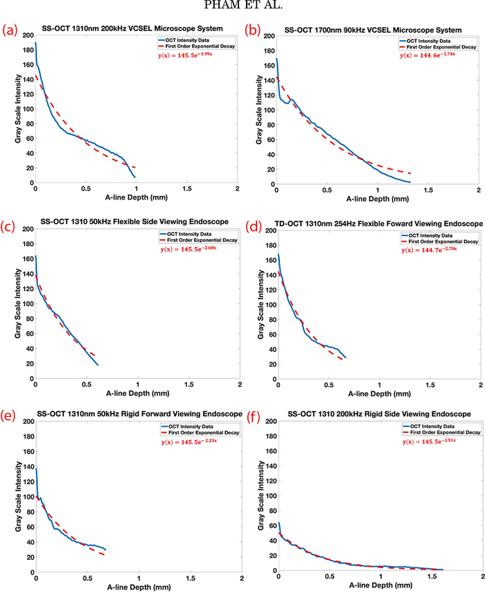
Fig. 7.
Average intensity based exponential decay curves and alpha constant of vocal folds imaged by each OCT system. Laterally averaged depth intensity and its exponential decay curves for the respective OCT systems and probes imaging the same fixed porcine larynx (a) SS-OCT 1310nm 200kHz VCSEL microscope system, (b) SS-OCT 1700nm 90kHz microscope system, (c) SS-OCT 1310nm 50kHz flexible side viewing endoscope, (d) TD-OCT 1310nm 254kHz flexible forward viewing endoscope, (e) SS-OCT 1310nm 50kHz rigid forward viewing endoscope, (f) SS-OCT 1310nm 200kHz rigid forward viewing endoscope. SS-OCT=swept-source OCT, VCSEL= vertical-cavity surface-emitting laser, TD-OCT=time-domain OCT.

