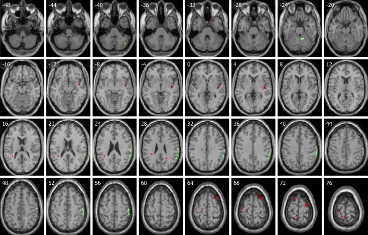
Figure 1.
Compared with baseline levels, idiopathic tinnitus patients before treatment showed increased activities in the right parahippocampa gyrus, right superior temporal gyrus, right superior frontal gyrus, anterior insula, left inferior parietal lobule, and left precentral gyrus. Decreased activities were in left postcentral gyrus and left ITG. (Red shows increased activity, and green shows decreased activity.)

