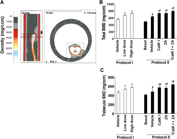
Fig. 5.
CatK inhibitor protects bone mineral density in mouse model. a Slices that were scanned by pQCT. The small box in the right panel indicates the tibia. The reference slice and the slices examined are indicated on a representative tibia. b Total bone mineral density detected by pQCT of tibiae from each group. c Trabecular bone mineral density detected by pQCT of tibiae from each group. *P < 0.01 compared to vehicle group; #P < 0.01 compared to the basal group

