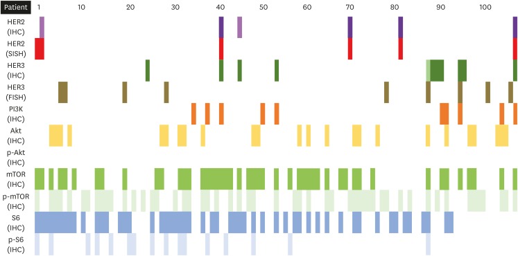
Fig. 1. Expression of all markers. In IHC of HER2 protein, 3+ and 1+ were marked with dark violet and violet, respectively. Dark green and bright green were used to show 3+ and 2+ on IHC of HER3 protein. In other markers, variable colours were used for protein expression or gene amplification. Empty slots are for negative findings.
FISH, fluorescence in situ hybridization; HER, human epidermal growth factor receptor; IHC, immunohistochemistry; SISH, silver in situ hybridization.

