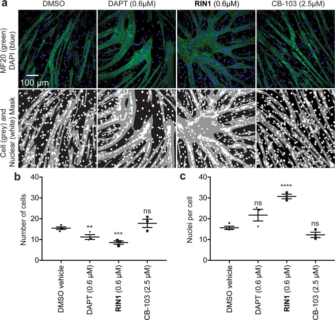
Figure 3.
Effect of RIN1 on C2C12 myoblast differentiation. Structured illumination photomicrographs of C2C12 cells at 4 days under permissive differentiation conditions and drug treatment as indicated. Upper panels: Cells were stained for myosin heavy chain with the MF20 antibody (green) and labeled with DAPI to identify nuclei (blue) (upper panels). Lower panels: Cell body and nuclei image masks for quantification. (b,c) Image analysis, n = 3 wells, quantified the number of cell mask (green) objects (b) and the ratio of nuclei per cell mask object (c). Assay repeated 3 times.

