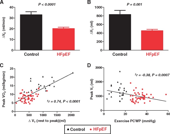
Figure 4.
(A) Compared with controls, subjects with heart failure with preserved ejection fraction displayed less increase in VE during peak exercise. (B) This was explained by impaired increase in VT in heart failure with preserved ejection fraction. (C and D) The change in VT varied directly with peak oxygen consumption, and peak VT was correlated inversely with peak pulmonary capillary wedge pressure. Error bars indicate SE. aDetermined by Pearson’s correlation analysis.

