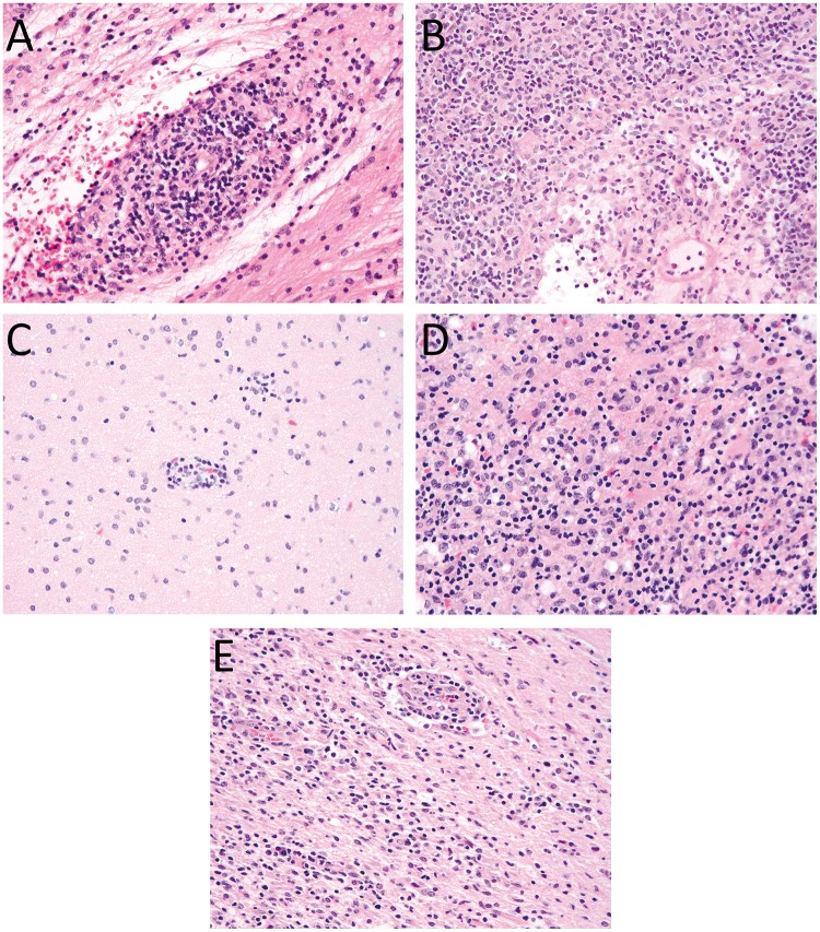
FIGURE 1.
Histological features present in brain biopsies. (A) Biopsy of the cerebellum from Patient 1 reveals a moderate perivascular, predominantly lymphocytic infiltrate. Lymphocytes infiltrate into small blood vessel walls causing some structural alteration and are associated with surrounding parenchymal edema. (B) Biopsy of the thalamus from Patient 1 shows a dense, predominantly lymphocytic perivascular and parenchymal inflammatory infiltrate with some structural alteration of the blood vessel walls and surrounding edema. (C) The initial frontal lobe biopsy from Patient 2 shows mild focal perivascular inflammatory infiltrates. (D) Frontal lobe biopsy of Patient 2 in the setting of recurrent disease shows severe destructive parenchymal lesions. (E) Similar diffuse parenchymal chronic inflammatory infiltrates are present in the frontal lobe biopsy of Patient 3. All images are stained with H&E and were taken with a 40× objective.

