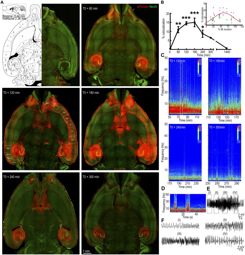
Figure 1.
The pattern of neuronal activation during status epilepticus. (A) Horizontal brain sections from animals injected with 4-OHT at different times from the start of continuous hippocampal stimulation (T0). A plate from the Paxinos mouse atlas at the level of the displayed sections is shown for comparison. The tdTomato fluorescence (red) corresponds to TRAPed cells and NeuN immunoreactivity (green) labels the neurons. Regions containing tdTomato-labelled neurons are marked as follows: AON = anterior olfactory nucleus; Pir = piriform cortex; AIV and AID = ventral and dorsal parts of agranular insular cortex, respectively; DI = dysgranular insular cortex; GI = granular insular cortex; Ect = ectorhinal cortex; PRh = perirhinal cortex; DLEnt, MEnt and CEnt = dorsolateral, medial and caudomedial entorhinal cortex, respectively. (B) The tdTomato voxels co-localized with NeuN immunoreactivity are expressed relative to that at T0. The values represent mean ± SEM of co-localized voxels from three consecutive 200-µm thick slices for each animal; n = 5 for T0 + 60 and n = 3 for T0, T0 + 120, +180, +240, +300 min and +24 h. *P < 0.05, **P < 0.005, ***P < 0.0005 versus T0, ANOVA with post hoc Dunnett’s multiple comparison test. The inset shows the co-localized voxels plotted against the time of 4-OHT administration normalized to the total duration of status epilepticus in each animal. The voxels from each of the three sections used for quantification are plotted separately in three colours. (C) Spectrograms show the power of the EEGs recorded from the hippocampal electrodes during 90 min prior to 4-OHT administration. This activity corresponds to the neuronal TRAPing captured in the images in A. (D) Power spectrum magnified from the boxed region in C, which corresponds to a tonic-clonic seizure. (E) A high amplitude fast spike-wave discharge on EEG corresponding to the band of high power marked by the box in D. (F) Magnified EEG, corresponding to regions (I), (II), (III) and (IV) in E. SE = status epilepticus.

