Figure 3.
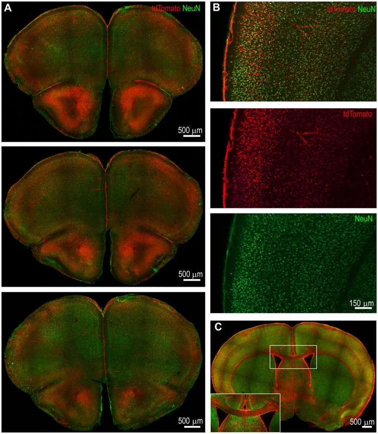
Neurons in the superficial layers of the motor and somatosensory cortex were TRAPed at the peak of status epilepticus. (A) Coronal sections from an animal at bregma 2.57 mm, 2.45 mm, and 1.97 mm (top to bottom) illustrate bilateral neuronal activation in the motor cortex at T0 + 120 min. The activation was more robust in rostral sections than in caudal sections. (B) Neuronal TRAPing in the somatosensory cortex at T0 + 120 min in a section at the level of bregma 0.01 mm. There was intense tdTomato expression in the superficial layers compared to the deep layers. (C) A coronal section at T0 + 120 min illustrating bilateral cortical activation and prominent labelling of the corpus callosum. The corpus callosum is magnified in the inset and shows tdTomato-filled axons, which originate in layer II/III of the cortex.

