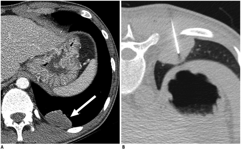
Fig. 2. Axial CT images in 21-year-old man diagnosed with acquired immunodeficiency syndrome.
He was referred for incidentally detected pulmonary mass during treatment of Pneumocystis jiroveci pneumonia.
A. Contrast-enhanced chest CT image shows 4.0-cm mildly enhanced mass in left lower lobe (arrow). B. CT-guided PTNB was performed by using 22-gauge aspiration needle, and pathologic examination showed chronic inflammation (false-negative result). Patient underwent left lower lobectomy, and lesion was confirmed to be malignant lymphoma in pathologic examination.

