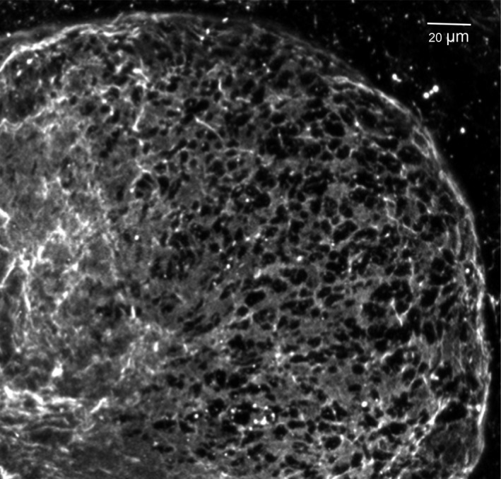
Figure 9.

Perforated basement membrane meshwork at a bud tip. This image shows an embryonic salivary gland bud that was expanding towards the right with its basement membrane stained for collagen IV (light grey). Note the intact basement membrane on the left that becomes perforated by numerous microscopic holes (black) towards the righthand tip of an expanding bud
