Figure 2.

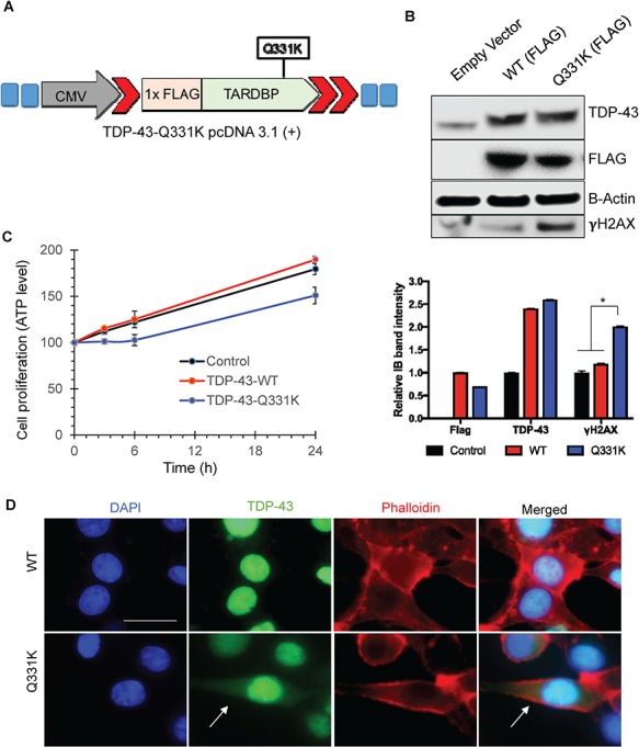
Ectopic transient expression of TDP-43-Q331K shows increased cytoplasmic accumulation in SH-SY5Y cells. (A) Vector construct expressing 1× FLAG and TDP-43-Q331K mutation. (B) Immunoblots of extracts from transiently transfected cells at 48 h, probed with FLAG, TDP-43 and γH2AX antibodies, show increased γH2AX, in cells expressing Q331K. β-Actin is shown as a loading control. The histogram shows quantitation of band intensity from three independent experiments. *P<0.01. (C) Cell-proliferation assay reveals reduced cell proliferation in Q331K-expressing cells. (D) IF of Q331K cells shows increased cytosolic accumulation of TDP-43 in cells expressing the Q331K mutant compared to WT. Left panel shows DAPI staining. Second from the left panel shows TDP-43 staining. Third panel shows Phalloidin staining as a cytoplasmic marker. Right panel show the merged image. Arrows indicate cytoplasmic localization of TDP-43 in Q331K-expressing cells. Scale bar represents 10 μm.
