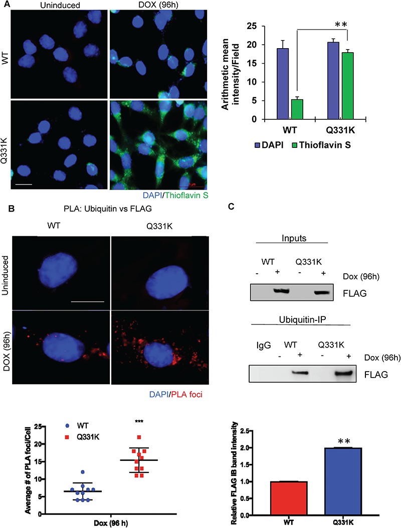
Figure 4.

Increased poly-ubiquitination and aggregation of Q331K in neuronal cytosol. (A) IF with Thioflavin S shows increased formation of aggregates in the cytoplasm of Q331K-expressing differentiated neurons (Scale bar, 5 μm). Histogram indicates quantitation of Thioflavin S aggregates. (B) Detection of PLA foci between ubiquitin and FLAG antibodies and quantitation of PLA foci from 25 cells (Scale bar, 10 μm). (C) Immunoblots of ubiquitin Co-IP from neuronal cells expressing TDP-43-Q331K, using FLAG antibody, show increased ubiquitinated TDP-43-Q331K compared to WT. Histogram represents quantitation of mean band intensity from three independent experiments. **P<0.05; ***P<0.0005.
