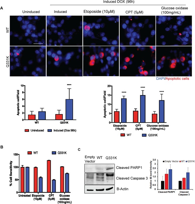
Figure 8.

Apoptosis and sensitivity of Q331K neurons to DNA damage. (A) TUNEL analysis by IF microscopy. Upper panels represent differentiated SH-SY5Y cells expressing WT while lower panels represent cells expressing Q331K mutation. Cells were induced with Dox for 24 h and cultured for an additional 72 h before treating with DNA damage-inducing drugs. The cells were then allowed to recover for 24 h before TUNEL analysis. Red staining indicates TUNEL-positive cells. Histogram represents the quantitation of TUNEL-positive cells per field from three independent experiments. (B) MTT assay for measuring the viability of cells after treatment with DNA-damaging agents. The viability was measured at 24 h of recovery after damage induction. (C) Immunoblots of cell extracts were probed for apoptotic markers with antibodies against cleaved PARP1 and cleaved Caspase 3. β-Actin is shown as a loading control.****P<0.0001.
