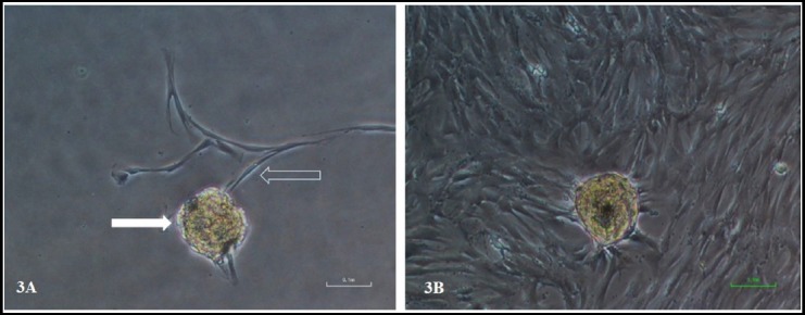
Fig. 3.

Dental pulp stem cells emerging from tissue explant. (3A) The micrograph showed the dental pulp tissue explant (solid arrow) and newly emerging cells from tissue explant (hollow arrow) at day 11 of culture. The cells exhibit typical morphology of fibroblast like cells with long cytoplasmic extension. (3B) showed same tissue explant after 19 days of culture with confluent field of view. Magnification 100X.
