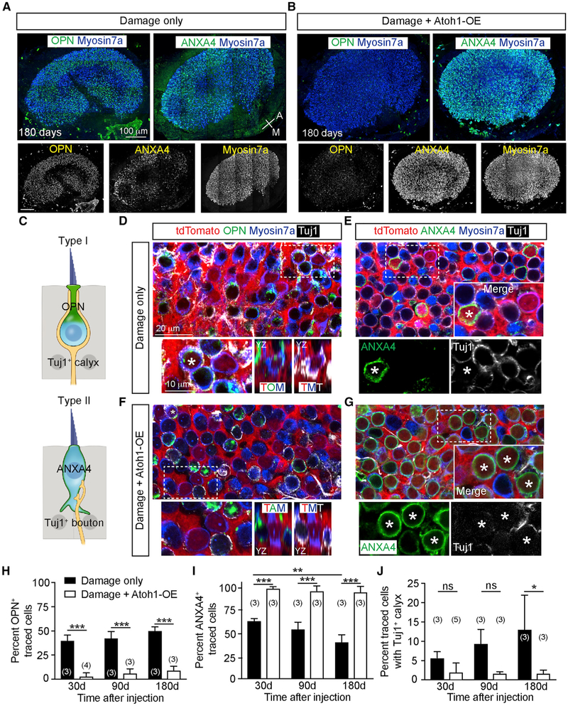
Figure 4. Atoh1 OE Increases Regeneration of Neurally Integrated Type II HCs.
(A and B) Damaged-only (A) and Atoh1 OE, damaged (B) utricles demonstrating osteopontin+ (OPN) type I HCs and annexin A4+ (ANXA4) type II HCs. After damage and Atoh1 OE, the utricle was densely populated by HCs, with a noticeable loss of OPN expression.
(C) Diagram depicting amphora-shaped type I HCs expressing OPN in the neck region and goblet-shaped type II HCs expressing membranous ANXA4.
(D) Representative images of damaged utricles at 180 days showing traced, Myosin7a+, OPN+ type I HCs (asterisk) with Tuj1+ calyx (orthogonal view highlighting OPN and Tuj1 labeling). Traced HCs negative for OPN were also noted.
(E) Many traced Myosin7a+, ANXA4+ type II HCs (asterisk) without Tuj1+ calyces were found, alongside traced HCs negative for ANXA4.
(F) After damage and Atoh1 OE, there was a noticeable loss of OPN expression and Tuj1+ calyces among traced Myosin7a+ HCs (orthogonal view illustrating loss of OPN labeling). A rare traced Myosin7a+, OPN+ type I HC (asterisk) identified with a Tuj1+ calyx.
(G) Most traced Myosin7a+ HCs expressed ANXA4 without Tuj1+ calyces. High-magnification images showing traced HCs expressing ANXA4+ (asterisks).
(H–J) Quantification of traced HCs expressing OPN (H), ANXA4 (I), and displaying calyces (J).
Two-way ANOVA with Tukey’s multiple comparisons test, n for utricles. *p < 0.05; **p < 0.01, ***p < 0.001. Data are shown as mean ± SD.

