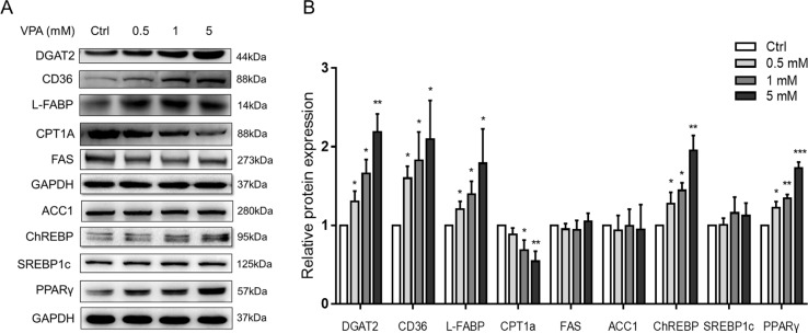
Figure 7.
Protein expressions of lipid metabolism genes following treatment with VPA. (A) L02 cells were treated for 24 h with VPA (0, 0.5, 1, and 5 mM). Whole-cell extracts were harvested and the expression levels of indicated genes and the internal control (GAPDH) were analyzed by Western blot. A representative blot is shown. (B) Band densities were quantified using ImageJ Software; band density of control groups was set at 1.00. Values are presented as mean ± SD (n = 3); *P < 0.05, **P < 0.01, ***P < 0.001 compared with the control group.

