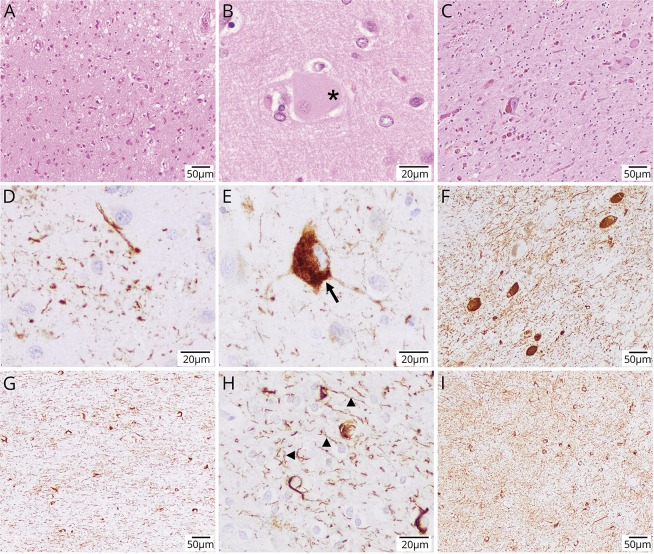
Figure 2. These images showcase the pertinent neuropathologic findings of case 2.
Hematoxylin and eosin staining shows superficial spongiosis in the postcentral region (A), a large achromatic or ballooned cell (highlighted by asterisk in B), and prominent nigral degeneration with severe neuronal loss and abundant extracellular neuromelanin pigment (C). (D–I) Abnormal pTau (AT8) and 4-repeat-tau-positive protein deposition on immunohistochemistry. Notable abnormal histopathologic findings included astrocytic plaques (D), frequent pretangles with some focal cytoplasmic condensations (E), tangles, pretangles, and abundant threads in the substantia nigra (F), very abundant threads (arrow heads) and coiled bodies (arrow) in the white matter (G and H), and abundant threads and pretangles in the striatum (I), overall consistent with the neuropathologic findings observed in corticobasal degeneration. Magnification scale bars are indicated in the bottom right corner of each panel.

