Figure 3.
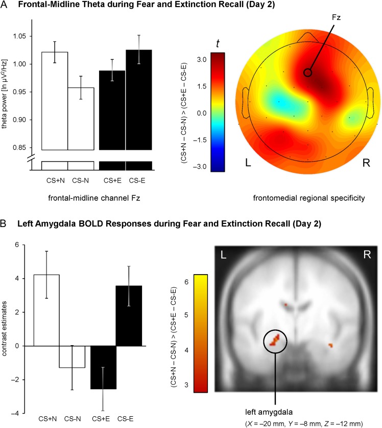
EEG and fMRI correlates of fear and extinction recall on Day 2. (A) Differential (CS+ – CS−) ln-transformed theta power at frontal-midline channel Fz was significantly reduced for extinguished versus nonextinguished stimuli (left). This effect was specific for frontomedial electrode channels (right). Bar graphs show the mean theta power (± within-subject SEM, O'Brien and Cousineau 2014). (B) Reduced differential amygdala responses (CS+ – CS−) for extinguished compared with nonextinguished stimuli. Habituation of amygdala activity was modeled by an exponentially decaying function, based on habituation of SCRs. For illustration purposes, the intensity threshold was set to P ≤ 0.005 (uncorrected) with a minimal cluster threshold of k ≥ 5 contiguous significant voxels. Activations (t-values) were superimposed on the MNI305 T1 template. All coordinates (X, Y, Z) are given in MNI space. L = left, R = right brain hemisphere. Bar graphs show the mean contrast estimates (± within-subject SEM, O'Brien and Cousineau 2014) for a cluster of voxels with P ≤ 0.005 (uncorrected) surrounding the peak voxel within the amygdala ROI.

