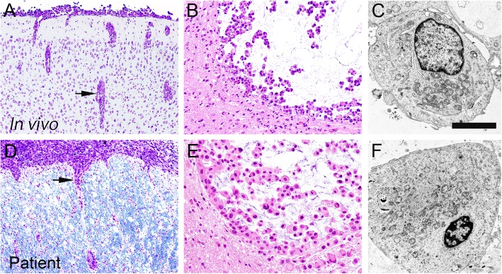
Fig. 2.
Morphological similarity of the tumor cells between those in the orthotopic xenograft (a, b, c) and those from the patient (d, e, f). The orthotopic xenografts share strikingly similar characteristics to those of the autopsy specimen, including the mode of tumor infiltration (a, d) and the light microscopic (b, e) and ultrastructural (c, f) morphology of the tumor cells. Scale bars a, d: 200 μm; b, e: 50 μm; c, F: 2 μm

