Fig. 1.
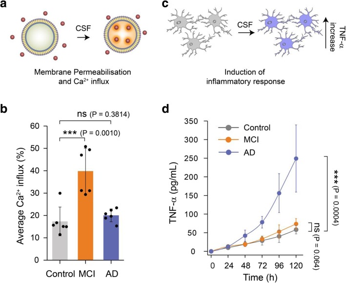
Soluble aggregates present in MCI and AD CSF samples show different dominant mechanisms of toxicity. a Membrane permeabilisation was measured by immobilising hundreds of vesicles containing a Ca2+-sensitive dye on PEGylated glass cover slides. If any species present in the CSF disrupts the integrity of the lipid membrane of the vesicles, Ca2+ ions from the surrounding buffer enter into individual vesicles in numbers that can be quantified using highly sensitive TIRF microscopy. b Aliquots of MCI CSF can cause more membrane permeabilisation compared to AD and control CSF (n = 6 AD, 6 MCI, 6 control CSF). A two-sample unpaired t-test was performed to compare each data set. c The inflammatory response in microglia cells was quantified using an ELISA assay to measure the levels of secreted tumour necrosis factor alpha (TNF-ɑ). d For this study, CSF samples were added to BV2 microglia cells and incubated for 120 h. Every 24 h the TNF-α concentration in the supernatant was quantified using an ELISA assay. AD CSF samples were more effective MCI and control CSF samples in inducing an inflammatory response (lines are guide to the eye; n = 10 AD, 6 MCI, 6 control CSF). Error bars are the standard deviation among data points. A two-sample unpaired t-test at the 120 h time point was performed to compare the data sets

