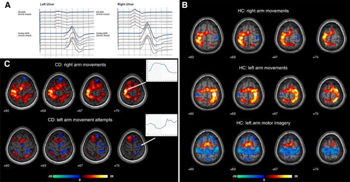
Figure 1.

(A) Motor evoked potentials in the conversion disorder (CD) patient for the paralyzed left and the healthy right ulnar nerve (recording site: abductor digiti minimi muscle [ADM]). The motor evoked potential latency and amplitude were within normal ranges without significant differences between both sides. (B) Task‐related activations (red–yellow) and deactivations (green–blue) in the healthy controls (HC) while performing normal arm movements and motor imagery. During normal motor execution deactivations were restricted to the primary motor cortex ipsilateral to the movement while during motor imagery widespread deactivations in the bilateral primary sensorimotor cortex were found. (C) Task‐related activations and deactivations in the CD patient and mean time course averaged over all blocks and runs (activation period indicated by gray background color) calculated for a 4 mm sphere centered on the activation peak (right arm movements) and deactivation peak (left arm movements) in the primary motor cortex in the CD patient. Movements of the healthy right arm elicited strong activations in the respective primary motor cortex with a signal increase during movement blocks (indicated by gray background color) for the activation peak (arrow). During movement attempts of the paralyzed left arm, the patient showed deactivation of the respective primary motor cortex further illustrated as signal drop during the motor task. [Color figure can be viewed at wileyonlinelibrary.com]
