
Figure 1.
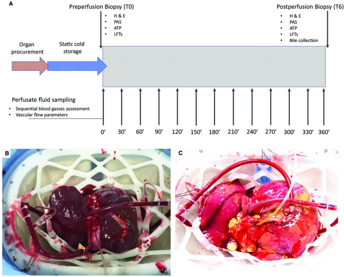
Study design and macroscopic appearance of a viable and nonviable liver. (A) The details of the study design and the perfusate fluid and biopsy sampling protocol. (B) A well‐perfused liver with optimal macroscopic appearance. The organ was rejected for transplantation due to the incidental discovery of a malignant melanoma. The liver began to function shortly after commencing the perfusion, and the vascular flows and blood gas profile patterns were used to help define criteria for liver graft viability (perfusion number 8). (C) A steatotic liver with suboptimal macroscopic appearance; this organ did not meet the viability criteria (perfusion number 2).