Figure 1.

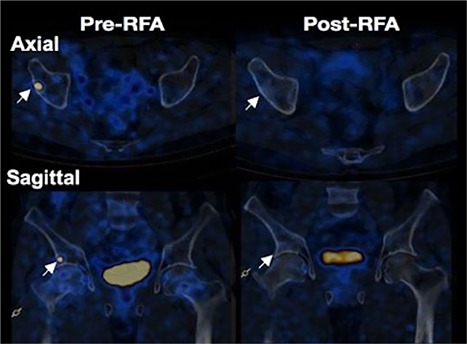
Pre‐ and post‐RFA Ga68 DOTANOC PET/CT scans of patient 1. Axial and sagittal views of the lesion in patient 1. Post‐RFA images demonstrate disappearance of the uptake at the lesion site. Arrows indicate the lesion.

Pre‐ and post‐RFA Ga68 DOTANOC PET/CT scans of patient 1. Axial and sagittal views of the lesion in patient 1. Post‐RFA images demonstrate disappearance of the uptake at the lesion site. Arrows indicate the lesion.