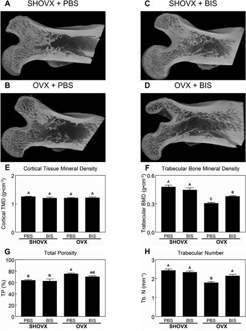
Figure 3.

Characterization of the Ovariectomy Induced Osteoporotic Phenotype. 8 month old, female, virgin, CD Sprague Dawley rats underwent sham ovariectomy (SHOVX) or ovariectomy (OVX) surgery. After 5 weeks, animals were injected with either ibandronate (BIS) or phosphate buffered saline (PBS) at a concentration of 25 μg/kg/25days followed by insertion of a Ti SLA implant 1 week later. After 28d of osseointegration, femurs were isolated and placed in 10% formalin. Femoral heads of the animals were analyzed with 3D microCT reconstructions (A–D). Cortical tissue mineral density (E), trabecular bone mineral density (F), total porosity (G), and trabecular number (H) were quantified from the microCT reconstructions. Data shown are the mean ± standard error (SE) of ten (n = 10) independent samples. Groups not sharing a letter are statistically significant at an α = 0.05.
