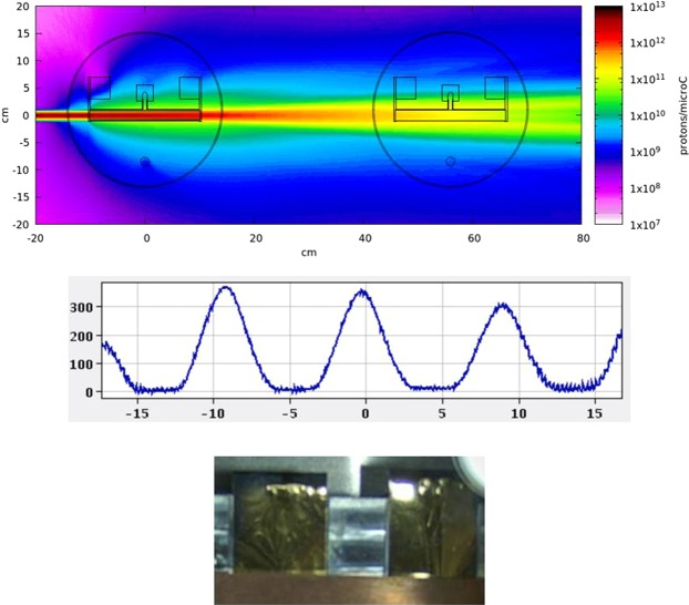
Figure 6.
Top: Fluka simulation25,26 showing the incoming proton beam on an ISOLDE target (3.5 g/cm2 UCx for the purpose of the simulation) and intercepting the MEDICIS target downstream. Middle: Screenshot taken with the beam scanner, located before the implantation chamber. Beams at A/q = 154,155,156 are seen (153, 157 partly visible). The collected beam is centred on A/q = 155, while isotopes present at other masses are physically removed from the implantation using mechanical slits located ahead of the foil. The horizontal scale is in mm. Bottom: Two zinc-coated gold foils in the collection chamber seen from the rear. The collection takes place on the foil located on the left.

