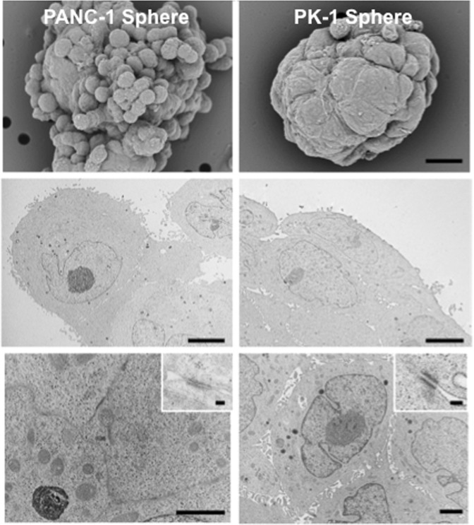
Figure 3.
Scanning electron microscopy (SEM) and transmission electron microscopy (TEM) analyses of PANC-1 and PK-1 spheres. SEM analysis showed that PANC-1 spheres were grape-like in appearance and that some cells had protrusions on their surfaces. SEM analysis of PK-1 spheres showed that they were round-to-oval with smooth cell surfaces. TEM analysis revealed the rough cell surface of PANC-1 cells and the lining cells of PK-1 cell spheres. Prominent adherent junctions and desmosomes were observed in PK-1 cells. Microvilli and secretory granules were observed in PK-1 cells. Scale bar: SEM = 10 μm, TEM middle and lower panels = 5 μm, TEM inset = 100 nm.

