Fig. 1.
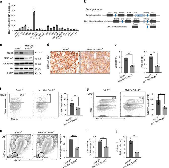
Setd2-deficient mice exhibit reduced numbers of T and B lymphocytes. a Relative mRNA expression of Setd2 in different hematopoietic cell compartments (n = 4). b Schematic of the strategy for establishing the Setd2f/f mouse line. c Immunoblotting of Setd2 and H3K36me3 in bone marrow nucleated cells (BMNCs) from Setd2 knockout mice. H3 and β-actin were used as the loading controls. d Immunohistochemistry of H3K36me3 in femur sections from Setd2 knockout and control mice. e Complete blood count of peripheral blood showed lymphopenia in pIpC-treated Mx1-Cre+;Setd2f/f mice. (WBC, white blood cell; PBL, peripheral blood lymphocyte; n = 7). f, g Flow cytometric analysis of CD3e+ T cells (f) and B220+ B cells (g) in the peripheral blood of Setd2 knockout and control mice (n = 8–9). h–j Flow cytometric analysis of bone marrow and bone marrow lymphocytes (BMLYs) in Setd2 knockout and control mice. (n = 6). (Data were collected at 8 weeks after the final pIpC injection. The data are presented as the means ± SDs. *p < 0.05; **p < 0.01; ***p < 0.001; ns, not significant, Source data are provided as a Source Data file)

