Figure 7:
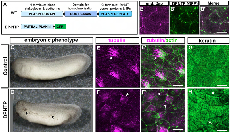
DP-NTP expression in X. laevis embryos. A) Schematic of the major domains of desmoplakin in wildtype (WT) and DP-NTP contruct. B) Desmoplakin labeled with an antibody B’) DP-NTP-GFP expression B”) Merge of B and B’. C,D) Lateral views of representative control (C) and DP-NTP expressing embryos. E-E’) Trunk epidermis of a representative control embryo, labeled with tubulin (pink, E) and phalloidin (green) merged with tubulin (E’). F-F’) Trunk epidermis of a representative DP-NTP expressing embryo, labeled with tubulin (pink, F) and phalloidin (green) merged with tubulin (F’). G,H). Keratin localization in the trunk epidermis of a representative control (G) DP-NTP expressing embryo (H). Scale bars =25 μm.

