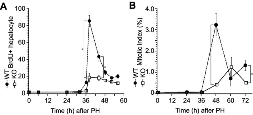
Figure 2.
Impaired hepatocyte proliferation after liver resection in the ERα knockout (KO) mice. Wild-type (WT) and KO mice underwent partial hepatectomy (PH), and the rate of hepatocyte proliferation was assessed for up to 72 h after PH. (A) Bromodeoxyuridine (BrdU) was injected 2 h before dissection. BrdU-positive hepatocytes were counted by immunocytochemistry. (B) Mitotic bodies were counted in liver sections. Values are expressed as the mean ± SEM (n=5–17 per group); *p<0.05.

