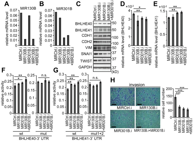
Figure 6. Inhibition of MIR130B and MIR301B enhanced the protein expression of BHLH40/41 and suppressed EMT in EC cells.
Expression levels of MIR130B (A) and MIR301B (B) in HEC-6 cells transfected with their inhibitors at a concentration of 50 nM. (C) Protein expression of BHLHE40/41 and EMT markers in HEC-6 cells used in (A, B). mRNA levels of BHLHE40 (D) and BHLHE41 (E) in HEC-6 cells used in (A–C). The reporter activities using 3’-UTRs of BHLHE40 (F) and BHLHE41 (G) in response to inhibitors of MIR130B and MIR301B are shown. The left graphs show the results from wild-type reporters and the right graphs show the results from mutant reporters (F, G). (H) In vitro cell invasion of HEC-6 cells used in (A–E). The right graph shows quantified results data (H). Data were representative from at least three experiments. The scale bars indicate 200 µm. MIRCtrl.i, control microRNA inhibitor; MIR130B.i, MIR130B inhibitor; MIR301B.i, MIR301B inhibitor; n.s., not significant; *, P < 0.05; **, P < 0.01; ***, P < 0.001.

