Fig. 1.
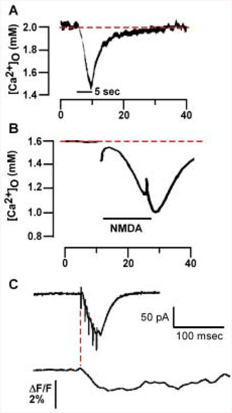
[Ca2+]o can drop following synaptic activity. A, Recordings with Ca2+-selective microelectrodes from guinea pig stratum pyramidale (SP) are shown during a 5-sec 10-Hz stimulus train in stratum radiatum (SR) (black bar). Adapted from Benninger and others (1980), Fig. 2, with permission from Elsevier Science. B, Recordings with Ca2+-selective microelectrodes from rat SR show a drop in [Ca]2+o following a 15-sec exposure to N-methyl-D-aspartate (NMDA) (black bar). Adapted from Heinemann and others (1990), Fig. 3, with permission from Elsevier Science. C, Evoked postsynaptic currents (EPSCs) and fluorescence measurements in SP following five 100-Hz stimuli applied to SR imaged using a cell-impermeant Ca2+-sensitive dye (Rhod-5N) and intracellular recordings show a decrease in Rhod-5N fluorescence following theta stimulation to SR. Note that the drop in fluorescence begins at the onset of the EPSC in SP (red line) and continues for several hundred milliseconds. Adapted from Rusakov and Fine (2003), Fig. 2, with permission from Cell Press. In both A and B, [Ca]2+o drops from baseline levels (dotted line) to 1.45 mM and less than 1 mM following either synaptic stimulation (A) or NMDA exposure (B), respectively.

