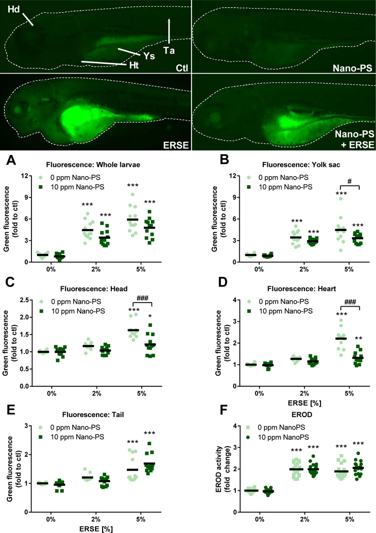
Figure 3:

EROD activity and total PAH bioaccumulation in zebrafish larvae exposed to 44 nm nanopolystyrene particles (Nano-PS) in the presence of a real world environmental PAH mixture (Elizabeth River Sediment Extract – ERSE). Animals were co-exposed at 6 hours post fertilization (hpf) to 10 ppm Nano-PS and 2% or 5% ERSE. The bioaccumulation was estimated by fluorescence microscopy at 96 hpf in the whole larvae (A) or the indicated areas (B – E), and EROD activity was assessed in vivo at 96 hpf. Representative fluorescence microscopy images are shown at the top panel and indicate the analyzed areas: whole larvae (dashed line), head (Hd), heart (Ht), yolk sac (Ys), and tail (Ta). Results are presented as scatter plot (n = 19 – 20 for EROD activity, n = 12 for fluorescence microscopy) and mean (black bar). Data were analyzed by two-way ANOVA followed by Tukey’s posthoc. Differences against the respective control group are shown as * (p < 0.05) and *** (p < 0.001), while differences between ERSE and ERSE + Nano-PS exposures are shown as # (p < 0.05) and ### (p < 0.001).
