
Figure 6. Histological assessment of local and distant tumors confirmed antitumor efficacy of the ADD-IT treatment protocol.
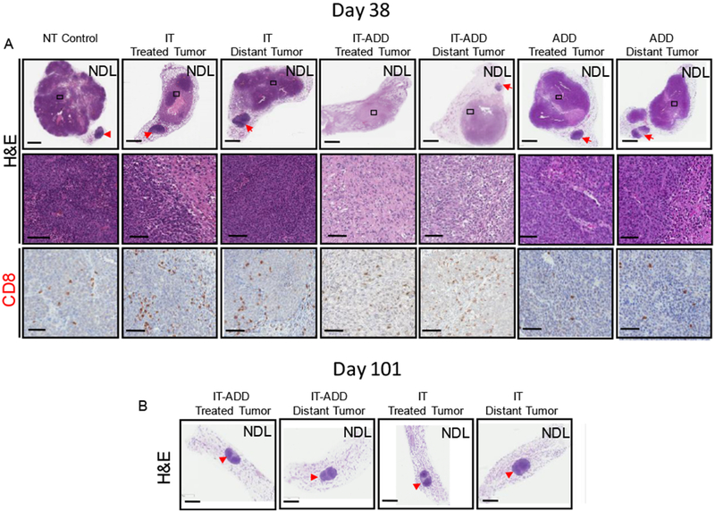
A) On day 38, a subset of mice was euthanized and tumors were isolated for histology and IHC. Histological sections stained for H&E (upper row, whole tumor view) and (middle row, magnified view), CD8 (lower row, magnified view). Whole tumor sections and the magnified views enclosed by black boxes are shown. B) H&E stained histological sections of mice treated with either IT-ADD or IT and survived 101 days. Red arrowheads denote the inguinal lymph nodes. Scale bars correspond to 3 mm (whole tumor panels) and 100 µm (magnified panels).