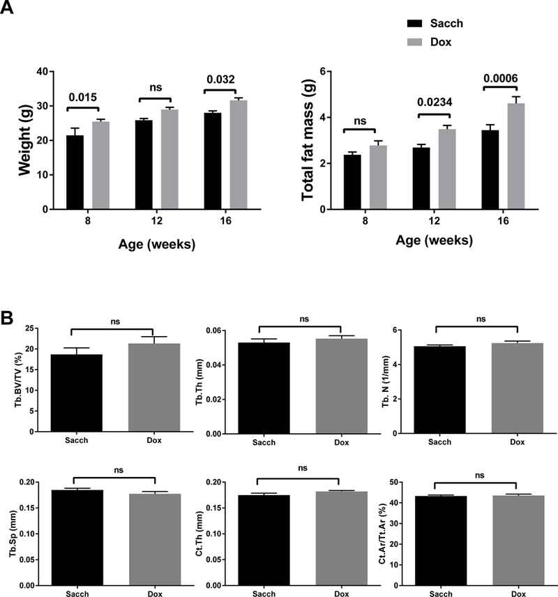
Figure 2: Dox treatment increases fat mass, but has no effect on bone microarchitecture in B6 mice.

Male mice were treated with either Sacch or Dox beginning at 4 weeks of age and followed till 16 weeks of age (12 weeks on Sacch or Dox). A) Body composition was determined by DXA at 8, 12, and 16 weeks of age (n=5–12); body weight and total fat mass are shown. B) Bone microarchitecture was analyzed in femur by μCT. No significant difference was observed in Tb. BV/TV, Tb.Th, Tb.N, Tb.Sp, Ct.Th, and Ct. Ar/Tt.Ar. Values represent mean ± SEM of n=6–9. p-values are calculated using GraphPad Prism as described in the methods section.
