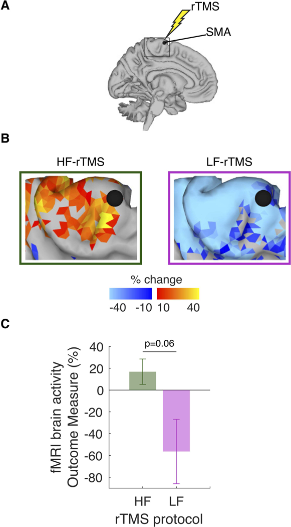
Figure 3. Effects of repetitive transcranial magnetic stimulation (rTMS) on resting brain activity around the representation of pelvic floor muscles (PFM) in supplementary motor area (SMA).

A. In each participant, high-frequency rTMS (HF-rTMS) and low-frequency rTMS (LF-rTMS) targeted the same PFM representation in SMA, with one-week wash-out period. B. Group resting-state functional magnetic resonance imaging (rs-fMRI) data. Brain maps of rTMS-induced changes, as assessed using fALFF in the 0.01–0.027 Hz frequency band before and after either HF-rTMS or LF rTMS. C. The effects of HF-rTMS and LF-rTMS on the resting SMA activity outcome measure trended toward statistical significance.
