Figure 2.
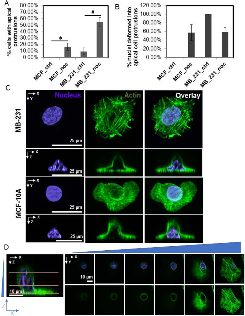
Nocodazole treatment enriches apical cell protrusions in MB-231 cells. A) Percentage of fixed cells with apical protrusions under various. Chi-square test was used to test statistical differences. *or #, p<0.05. N > 30 cells for each condition (MCF_ctrl: untreated MCF-10A cells; MB_231_ctrl: untreated MB-231 cells; MCF_noc and MB_231_noc: MCF-10A cells and MB-231 cells respectively treated with 5 μM nocodazole). Error bars indicate SEM. B) Percentage of cells with an apical cell protrusion in which the nuclear shape was vertically deformed into the protrusion. Error bars are SEM. C) Representative fluorescent images of an MB-231 and MCF-10A cell treated with nocodazole for 1 hr, showing the apical cell protrusion and associated vertical nuclear deformation. D) Fluorescent images show confocal images at different planes (marked by horizontal lines) of an MB-231 cell treated with nocodazole. The apical protrusion is present in an otherwise well-spread cell.

