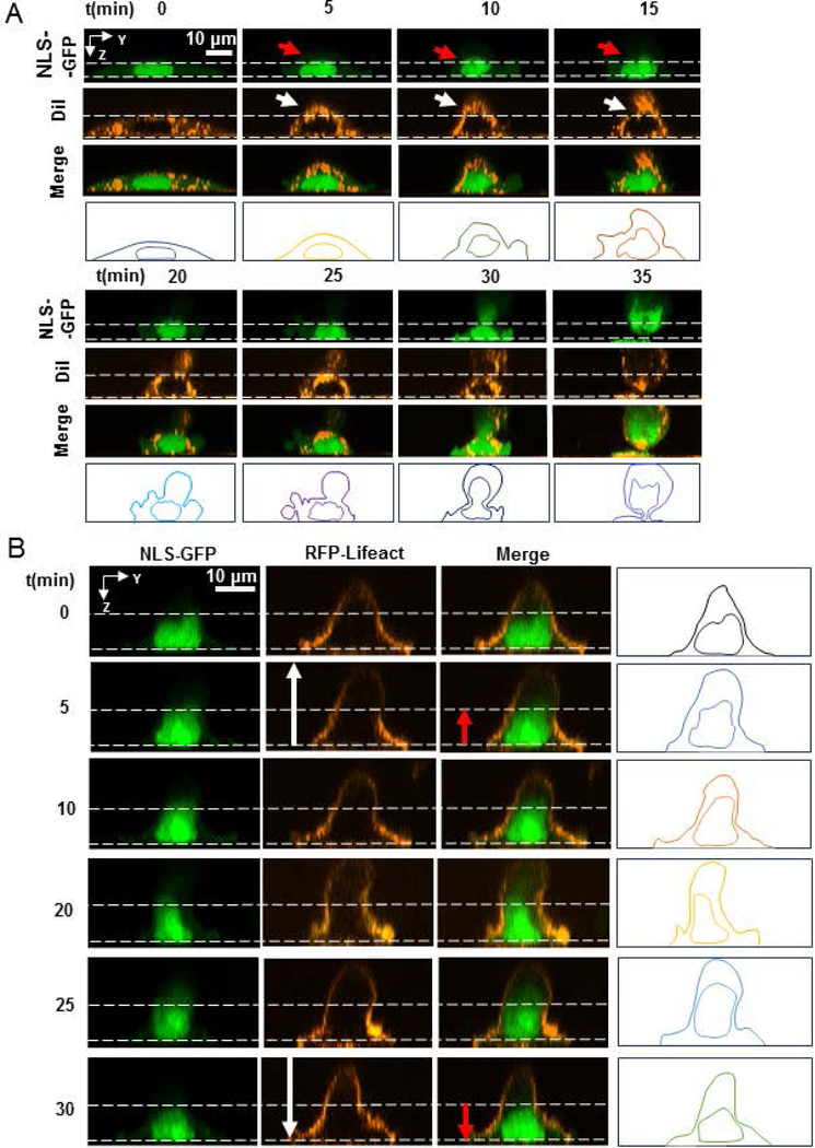
Figure 3.
Apical cells protrusion precedes vertical nuclear deformation. A) Reconstructed fluorescent time-lapse images of the vertical cross-section of a live cell expressing NLS-GFP and treated with lipophilic tracer DiI showing the formation of a vertical protrusion in the cell membrane near the nucleus (white arrows) and the consequent deformation of the nucleus (red arrows) after treatment with 5μM nocodazole at time t = 0 min. For reference, vertical dashed lines indicate the initial position of the nucleus and cell membrane. B) Reconstructed fluorescent time-lapse images of the vertical cross-section of a living cell expressing NLS-GFP and RFP-LifeAct showing the formation and retraction of a vertical cell protrusion near the nucleus (white arrows) and the consequent deformation of the nucleus (red arrows) after treatment with nocodazole at t = 0 min. Vertical dashed lines indicate the initial position of the nucleus for reference.

