Figure 4.
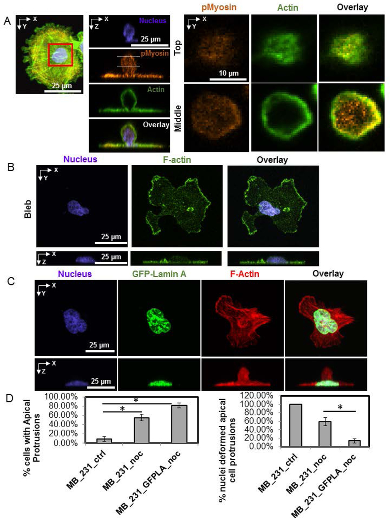
Apical cell protrusions are NMMII dependent, while vertical nuclear deformation is reduced upon over-expression of GFP-Lamin A. A) An MB-231 cell treated with nocodazole for 1 hr before fixation and stained for pMyosin (orange), F-actin (green) and nucleus (blue). Inset at two z-positions (top and middle planes) shows the distribution of F-actin and pMyosin in the apical cell protrusion. B) Fluorescent images show an example of a MB-231 cell treated with blebbistatin, which abrogated the apical cell protrusion. C) Images show a GFP-Lamin A expressing cell treated with nocodazole for 1 hr before fixation. An apical protrusion is clearly visible, but with no vertically upward deformation. D) Bar plot shows the frequency of cells with apical protrusions of MDA-MB-231 cells (left) and frequency of nuclei deformed into the apical cell protrusions. (MB_231_ctrl: untreated MB-231 cells; MB_231_noc: MB-231 cells treated with 5 μM nocodazole; MB_231_GFPLA_noc: MB-231 cells expressing GFP-Lamin A and treated with 5 μM nocodazole). Error bar is SEM. At least 32 samples from 3 different dishes are analyzed. Chi-square test with Bonferroni correction was used to test statistical differences. *, p<0.05.

