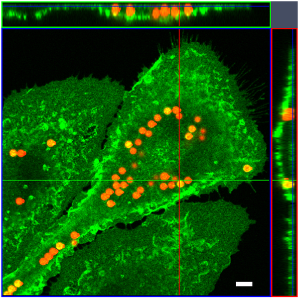
Fig. 1.
Fluorescent beads engulfed by an activated microglial cell. The z stack image taken on a Zeiss confocal microscope. Ortho image assembled from a Z-stack. The image shows XZ plane (green line) and YZ plane (red line) through the stack of images. Section of the XY plane (blue) is the slice plane of the stack. The result of an orthogonal section is visible at the image margins (above and right). Scale bar 5 μm.

