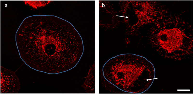
Fig. 4.
Morphological alterations were seen in stimulated cells in absence and presence of LA. The cells are outlined in blue to highlight difference in cell size. a. LPS/IFN-γ stimulated cells b. LPS/IFN-γ stimulated cells that were also treated with LA 25 μg/ml. Microglial cells were fixed with paraformaldehyde and stained with WGA after 24 hrs. Arrows indicate blebs. Scale bar 10 μm.

